It is well known that the human body cannot fully heal large-scale bone defects and, in most cases, requires external surgical intervention to return to normal. At present, the commonly used bone graft materials in clinical practice are as follows: autologous bone, that is, the material is taken from the patient's own body, which is the most ideal restoration, but there are secondary injuries, multiple operation areas and donor site complications, sources. Insufficient problems; allogeneic bones, mostly from corpse donations or animals, have immune responses, potential infection risks, and ethical issues.

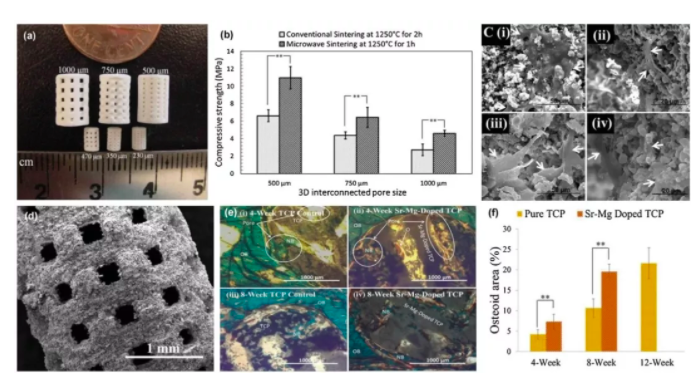
Figure 1: Mechanical properties and biological properties of 3D printed artificial bone scaffolds with different post-treatment processes and different compositions
In this context, 3D printed artificial bone scaffolds have become a research hotspot of relevant researchers. Because this technology enables the direct fabrication of porous scaffolds with design shapes, controlled chemistry, and interconnected pores, it has become the only choice for making bone repair materials. This article will introduce the latest developments, current challenges and future directions of 3D printed artificial bone scaffolds in a comprehensive article published in Materials Today's Bone tissue engineering using 3D printing.
The review first gives a background introduction. The bone tissue of the human body consists of two different structures: cancellous bone and cortical bone. The cancellous bone is spongy and has a porosity of 50 to 90%. Cortical bone is a dense outer layer of bone with a porosity of less than 10%. For continuous ingrowth of bone tissue, interconnected pores are very important to allow nutrients and molecules to be transported into the interior of the scaffold, promoting cell ingrowth, vascularization, and waste removal. Currently, porous bone scaffolds can be fabricated by a variety of methods, such as chemical/gas foaming, solvent casting, particle/salt leaching, freeze drying, and the like. However, these methods do not control the aperture, shape and interconnectivity. The design and manufacture of such a bracket using the 3D printing method can effectively solve the above problems.
The paper then concludes that the strength and degradation properties of the porous scaffold are greatly affected by the pore size, geometry and support direction with respect to the loading direction. Secondly, the surface properties of the material such as surface charge and morphology also affect the hydrophilicity of the material, which in turn affects Bone tissue ingrowth. The article goes on to point out that low mechanical strength is a major challenge in porous scaffolds, and that optimized post-treatment methods and compositional modifications can improve the mechanical properties of artificial bone scaffolds.

Figure 2: Example of 3D printed cranial structure and implant structure
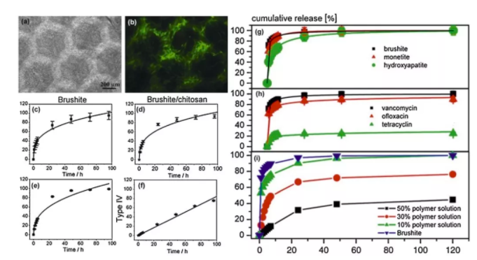
The use of 3D printed stents for growth factors and drug delivery is also a research hotspot. This method not only reduces the dose required for systemic delivery, but also greatly reduces side effects and also controls the release pattern of the drug. It is concluded that the size of the stent aperture, connectivity and geometry are effective parameters for controlling drug loading and rate of release in vivo.
The paper highlights the current challenges and future directions of 3D printed artificial bone technology. It is pointed out that the residue from the adhesive may be difficult to remove during the sintering process. The ability to achieve higher accuracy and resolution, and to make smaller apertures, depends on powder characteristics and build parameters. And 3D printed artificial bones always require post-treatment, such as sintering or densification at high temperatures. During the sintering process, the overall shrinkage is uneven, which causes a large amount of cracking of the part and renders it unusable. Due to this drawback, it is very difficult to simulate the structure in which the aforementioned cancellous bone and cortical bone coexist using 3D printing. Another post-processing challenge is to remove loose powder from the interconnected pores in the part. The residual powder in the hole may be sintered with the porous portion, making it less interconnected with the design part and further reducing the size of the hole after sintering. .

Figure 3: Drug release based on 3D printed artificial bone scaffold
The need for 3D printing technology in this area will continue to increase in the future due to the ability to tailor patient-specific defects and specific clinical needs. For artificial bone 3D printing, the most critical issue to be aware of is the mechanical properties of the porous scaffold. However, increasing the porosity will reduce the strength of the stent, but the use of degradable polymer infiltration to enhance the strength and toughness of these stents is one way to solve this problem. Second, printing live cells or adding growth factors/drugs will be another promising research direction. Finally, the article points out that although the current technology allows us to establish a structure similar to the organization, we still have a long way to go from the full print function organization. More process-property optimization, in vitro and in vivo studies are needed in this direction to continue to advance the development of bone tissue engineering.
Author: Li Xiang
Latex Mattress,Latex Spring Mattress,Latex Foam Mattress,Mesh Latex Foam Mattress
HESHAN HAIMA FURNITURE CO., LTD , https://www.springmattress.com